Vision exams are performed to determine the overall health of your eyes and to assess how well you can see. And as we age, they examinations are essential.
While the exact procedures vary by provider, your doctor may examine your eyes with special lights and perform an eye pressure test. Or, they might have you read from a chart on the wall to determine if you need corrective lenses. Your doctor may even place a unit in front of your face that will help determine your new prescription, if needed.
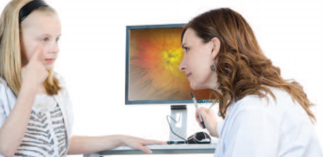
Retinal examinations are performed so doctors can see the back of your eye. This exam is performed so a doctor can see if you’re developing retinal disease or other serious problems that can affect your vision. During follow up exams, your doctor may compare your new results with past ones to detect any changes and begin treatment if necessary.
There are two methods for examining the retina. One way is to dilate the eyes with eye drops that keep your pupil open even when the doctor shines a bright light into your eye. This way, your doctor can see the back of your eye where the retina is located. The eye drops take about 10 minutes to become effective, and they can blur vision and cause light sensitivity for roughly several hours after the exam.
The other way to perform this exam is to use an optomap retinal imaging device. For this method, eye drops are not required and the exam can be done in mere minutes. All you have to do is simply place your eye on the face plate and the optomap retinal imaging device will scan each eye, providing your doctor with a high-quality image of your retina.
Both ways are perfectly acceptable and in many cases they are complementary.
Next time you’re advised to get a vision or retina examination, be sure to ask your doctor if they have optomap.
Image Source: optomap