At Optos, delivering breakthroughs in pediatric ocular health is a driving vision. Douglas Anderson founded Optos in 1992 with the express goal of developing an imaging technology that could provide early diagnosis of the kind of retinal detachment that ultimately caused his five-year old son, Leif to lose vision in one of his eyes.
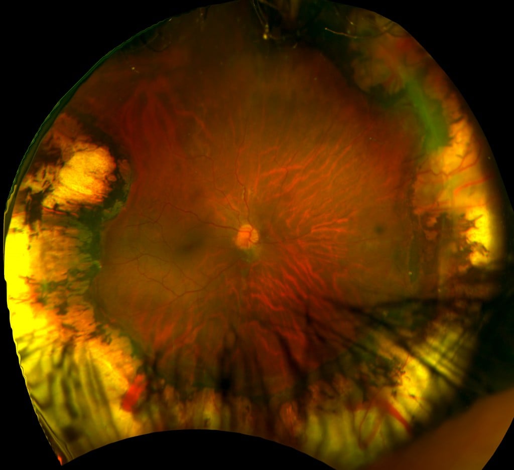
Retinopathy of Prematurity – Daytona
Now, almost a quarter-century later, UWF™ (ultra-widefield imaging) technology developed by Optos is transforming the way optometrists and ophthalmologists care for their patients. UWF imaging enables faster, more comprehensive routine exams, increases the cost/effectiveness of telehealth programs, improves the scope and accuracy of screening for diabetes and other eye disease, and makes possible new approaches to managing and tracking treatment. And, if you’re a pediatric ophthalmologist, UWF imaging is making possible new, better approaches to diagnosis and treatment.
Practitioners treating infants with ROP (retinopathy of prematurity) report1 that UWF imaging delivers clinically useful, high-quality post-treatment images while overcoming some of the problematic aspects of traditional imaging technology. The authors observed that a major advantage of ultra-widefield technology is that images can be obtained without placing the diagnostic instrument in direct contact with the infant’s eyes. This eliminates a potential source of post-operative infections. Non-contact UWF imaging — which is generally faster than traditional digital imaging — also reduced the need to anesthetize some infants so as to immobilize their eyes and/or limit head motion. Add to this UWF imaging’s wider coverage area, and the result is a safer, less-invasive and highly effective diagnostic technique.
Other complex diagnostic procedures are also being positively impacted by UWF imaging. In instances where infants must be examined using fundus fluorescein angiography, the required fluorescein is normally administered by IV, and the procedure is conducted in an operating theater or neonatal unit. A pioneering study2 reported on a case in which oral fluorescein was used to obtain high-value UWF optomap fa images from an infant with incontinentia pigmenti. The use of oral fluorescein allowed the entire procedure to be performed in an office setting, illustrating yet another way UWF imaging is enabling safer, less invasive diagnostic methods.
Other researchers are contributing to a growing body of pediatric UWF imaging practices. One retrospective study examining a group of older children (average age 9.3 years), evaluated the impact UWF fa in the diagnosis of such conditions as to uveitis, hereditary retinal dystrophies, retinal vascular diseases, infection and tumors2. Pathologies in the retinal periphery, which might otherwise have not been identified using traditional methods, were found in 75 percent of the cases. Another case study looked at the impact of UWF fa in treating and managing a small group of pediatric patients with Coats’ disease and familial exudative vitreoretinopathy (FEVR). The authors concluded:
Ultra-widefield fundus photography and UWFA can be used successfully as an outpatient procedure in the pediatric patient population without the necessity of examination under anesthesia and can aid the physician in the documentation and evaluation of peripheral retinal pathology. UWFA can also assist in directing laser photocoagulation in the treatment of pediatric retinal diseases3.
Important diagnostic procedures for infants and children made safer, less invasive, and more comprehensive.
But beyond this, UWF imaging can have a profound impact on something as simple as a follow-up exam. Imagine how a typical, high-energy 6-year-old reacts to a traditional dilation and slit-lamp examination, and then watch as the youngster in this video saunters up to an Optos Daytona, and, in moments, is looking at his complete color optomap.
Pediatric ophthalmologic practice is uniquely challenging, and UWF imaging is addressing many of these challenges in ways that will lead to more accurate diagnoses and better patient outcomes. Visit the r contact us directly.
Sources:
- Non-contact Ultra-widefield Imaging of Retinopathy of Prematurity Using the Optos Dual Wavelength Scanning Laser Ophthalmoscope. Eye. 2013.
- Pediatric retinal conditions imaged by ultra-wide field fluorescein. Ophthalmic surgery, Lasers and imaging Retina. 2013
- Ultra-widefieldimaging for the management of pediatric retinal diseases. Journal of Pediatric Ophthalmology and Strabismus. 2013.