As medical communities around the globe work to make the transition from treating sickness to preventing it, it’s still not the norm for healthy, asymptotic individuals to receive routine screening exams.
Unexpected Results When “Healthy” People are Examined with UWF Retinal Imaging
Ocular health is no exception. How often do adults with 20/20 vision and no eye-related symptoms schedule themselves for an optometric exam? That’s especially of concern considering that a number of ocular diseases such as, diabetic retinopathy (DR)1, open angle glaucoma2, age-related macular degeneration (AMD)3, and degenerative retinoschisis4, may not present any symptoms during their initial phases. Early detection of these diseases can have a significant impact on courses of treatment and the probability of positive outcomes.
One illustration of how UWF™ (ultra-widefield) imaging can improve the early detection of eye disease are the results of what amounts to an inadvertent experiment in the screening of healthy individuals. Training for Optos users and new Optos technical employees involves hands-on familiarization and instruction on UWF imaging systems. Part of that involves trainees taking color optomap images of themselves. These training exercises sometimes yield unexpected results:
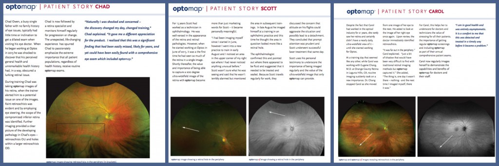
— Chad, who joined Optos in 2013, is a single parent and at the time of his hire had no history of eye problems. Prior to coming to Optos his full schedule and absence of symptoms had made getting an optometric exam a low priority. During training Chad’s instructor observed faint retinoschisis on one of Chad’s optomap images. Further examination using eye steering enabled Chad and his instructor to fully visualize the extent of Chad’s retinoschisis, which included holes in a larger area of retinoschisis in his right eye. Chad’s now receiving regular medical care and follow-up.
— Scott joined Optos in June 2015 and brought with him 15 years of experience as an ophthalmologic technician. His initial instruction included routine color optomap images of his own eyes. Then, in early August, he imaged himself during a training session and noticed an area in his upper right eye that he’d not seen on earlier images. Unsure of what this was, he kept track of it during other training sessions. In late August Scott imaged himself again during a training session at an ophthalmic practice and this time the suspect area had resolved itself into something identifiable – a retinal hole. The ophthalmologist participating in the training confirmed this and also observed related leakage. He conferred with Scott and discussed the possible risks associated with his frequent business travel. Scott received laser treatment later that same day.
— Carol, an optical industry professional with 27 years of experience, had never viewed her retina nor had an ultra-widefield examination until she started working for Optos. Then, during a training session with an MD, the doctor stopped Carol as they were viewing color optomap images of her eyes. Asking to look once more at the image of her right eye, the doctor identified an area of retinoschisis visible on the far periphery of the retina. Carol has received follow-up treatment, and makes a point during training sessions to image herself and to share her personal experience with the doctors and medical professionals with whom she works.
These stories are by no means unusual, and while not part of a statistically significant study, they make a simple, important statement about ocular healthcare – the wider use of ultra-widefield retinal imaging for routine screening has the potential to significantly improve personal health outcomes.
We always suggest that everyone include optomap as a part of their comprehensive eye exam. To find a provider near you, visit www.optomap.com.
Sources:
- https://nei.nih.gov/health/diabetic/retinopathy
- https://nei.nih.gov/health/glaucoma/glaucoma_facts
- https://nei.nih.gov/health/maculardegen/armd_facts
- https://www.institut-vision.org/en/health/eye-diseases/28-diseases/94-degenerative-retinoschisis.html?showall=1&limitstart=