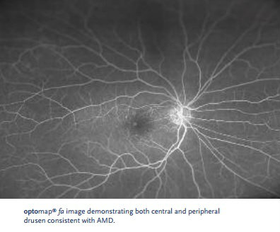
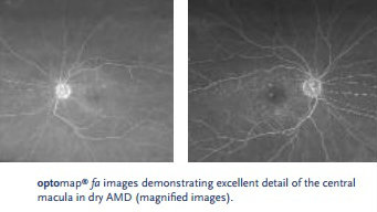
The Optos® ultra-widefield (UWF) scanning laser ophthalmoscope was first introduced at Duke University Eye Center as a part of its comprehensive eye care services in September 2006. Over the past seven years, the practice observed a number of benefits the device added to its services, including improved image quality and increased field of view. In one case of dry AMD, the technology helped illustrate “the unique value of (UWF) fluorescein angiography by Optos” in patients suffering from AMD.
A female patient was diagnosed with dry AMD 1.5 years earlier with nuclear sclerotic cataracts in both of her eyes. At a routine appointment, the exam revealed a change in the patient’s visual acuity since her previous exam six months earlier. Her visual acuity was now 20/100 in the right eye and 20/40 in the left, with an Intraocular Pressure (IOP) of 15 and 18 in the right and left eyes, respectively.
Practitioners used the UWF scanning laser ophthalmoscope from Optos to gather clear images of this patient’s retinas even though she had rather dense cataracts. This used to be a challenge with traditional equipment like mydriatic white-light cameras, however, with the red-green laser functionality, there was “less scatter as the beams enter the anterior segment allowing for a better image of the retinal surface.” The images gathered showcased how this patient’s case of AMD was affecting her retina in the central and peripheral regions. Practitioners were also able to confirm that the patient didn’t have wet AMD at the time of the appointment.


Providing the patient with a more comprehensive retinal exam and the assurance that she didn’t suffer from wet AMD is just one of the many benefits the team at Duke University Eye Center experienced since bringing Optos’ technology into their practice. The greater field of view – 200° in one image – has helped improve their ability to diagnose retinal diseases like dry AMD. As a result, the progression of the disease across the surface of the retina can be captured at each stage.
Are you interested in learning more about UWF retinal imaging technology from Optos, and how it can benefit your practice? Visit our website today to read more case studies and to learn how we can help you see more and treat more.