In several ocular diseases, the peripheral retina is the site of pathology. From conditions such as diabetic retinopathy to familial exudative vitreoretinopathy (FEVR), to retinal tears and detachments, the peripheral retina has been an area of interest for many eye care professionals throughout the ages.
Searching for a better means of treating conditions of the peripheral retina has been a challenge for researchers for well over a century, and during that time, advancements in imaging technology have allowed for easier evaluation by use of photography.
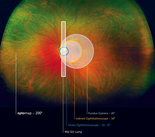
Listed below is an abbreviated history from the Review of Ophthalmology of how imaging the peripheral retina has evolved into the ultra-widefield imaging of today:
- — 1851- The first ophthalmoscope was invented by Hermann Von Hemholtz.
- — 1926- The first fundus camera that could provide a 20 degree view of the retina is invented, allowing ocular fundus structure documentation. Many years later, a camera with the capability of a 30 degree view would be the new standard. Although this allowed a view of the posterior pole and the optic nerve, periphery viewing was very limited.
- — In time, doctors were able to use a regular fundus camera to view up to a 75 degree field of view by taking photographs using seven standard fields and creating a montage. While this method was helpful in detecting diabetic retinopathy and sickle cell retinopathy, it still did not encompass enough of the periphery.
- — 1997- Technology was created that allowed a 130 degree view of the retina when the widest-angle lens was used. It consisted of a wide-angled camera, contact lens and multiple lens attachments. Shortly thereafter, SLO technology using laser light and scanning created high resolution photographs.
- — 2000- Optos introduces what is still the only ultra-widefield imaging technology. We are the only company to allow a 200 degree view of the retina which equates to approximately 82.5 percent, including into the periphery. A non-contact and non-invasive single scan of the patient’s eye produces a high quality digital image that not only provides an unprecedented view of the retina, it is also capable of color, red-free af and fa imaging.
Retinal imaging has a long and impressive history, and as the only clinically validated ultra-widefield imaging technology that aids clinicians to improve patient outcomes with the ability to “See More. Treat More Effectively. ™,” we invite you to contact Optos to learn how partnering with us can advance your practice.