The expanding role of optometrists as primary eye care providers is also increasing expectations about the level of diagnostic and follow-up care. Whether it’s screening and monitoring hypertensive or diabetic patients, diagnosing a patient’s vision impairment, or uncovering pathology during a routine examination, optometric practices are expected to know more and do more about the ocular health of their patients.
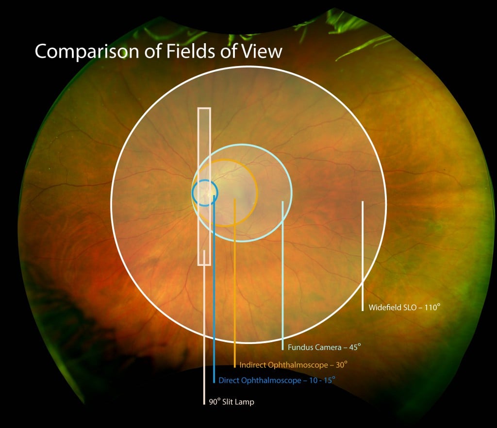
Comparison of Fields of View
Many optometrists may be meeting these expectations by utilizing ultra-widefield (UWF™) retinal imaging as a routine part of their practices. Along with improving patient experience and practice productivity, UWF retinal imaging gives these practitioners a powerful tool that is improving their practices’ overall optometric care.
UWF Retinal Imaging – A Versatile Imaging Platform
UWF retinal imaging provides practitioners with a versatile screening platform that can strengthen their ability to identify ocular and systemic disease while enabling more comprehensive and accurate treatment plan.
— One important capability offered by UWF retinal imaging is comprehensive and detailed visualization of the retinal periphery. As this technology proliferates throughout the eye care community, more studies are finding that pathologies of the retinal periphery are important to the understanding, diagnosis and treatment of a variety of conditions. One recent report showed that over a four-year period, diabetic patients with predominantly peripheral retinal lesions were 4.7 times more likely to progress to proliferative diabetic retinopathy1. This has implications for the screening and monitoring of diabetic patients.
— Other research work has demonstrated that UWF retinal imaging can characterize retinal nerve fiber layer (RNFL) defects that in turn correlate to findings by GDx. The authors observed that UWF imaging results of this kind provide a useful adjuvant in glaucoma screening.
— Diagnostic capture is another strength of UWF retinal imaging. As this individual case study suggests, UWF color images may detect pathologies that even experienced practitioners may miss during binocular indirect ophthalmoscopy. Further, the ability to manipulate images via zoom, contrast, and red/green channel separations adds to the practitioner’s ability to examine and diagnose suspect features.
— UWF imaging systems configured for optometry support both color and autofluorescence (AF) modalities. This gives the practitioner a visualization of metabolic changes in the retinal pigment epithelium (RPE) that in turn may guide further follow-up examinations and/or referrals.
— Whether in discussion with a patient or as part of a referral, UWF retinal images taken over a period of time can accurately track important retinal changes as well as the progression of pathologies. For patients, it’s a compelling narrative that reinforces the practitioner’s guidance, whether it be continued monitoring, ongoing treatment, or referral to another physician.
Other Practice Improvements
UWF retinal imaging can also assist in the improvement of other areas of the practice:
— It overcomes the difficulties in conducting dilated slit lamp examinations on children, and in so doing improves the quality of the examination (in this case study, uncovering retinal detachment and choroidal melanoma in a child otherwise reluctant to be examined).
— Referrals to other physicians, particularly ophthalmologists, is at once simplified and enhanced. Workflow software enables easy storage and transmittal, and the high-resolution images reduce the need for elaboration about the size and location of the suspect pathologies.
UWF retinal imaging improves optometric care by improving practitioners’ abilities to visualize, diagnose, track and manage their patients’ ocular health. More than a highly capable retinal imaging platform, its ease-of-use assures wide acceptance and utilization by both patients and staff.