Uveitis is an inflammatory condition that can occur anywhere in the eye and is associated with a number of diseases and conditions. These related illnesses make it difficult to estimate uveitis’ impact on global health, but studies suggest it could be responsible for as many as 10% of the cases of total blindness worldwide.
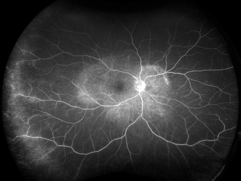
optomap fa, Uveitis Late OD, California – Courtesy of Retina Consultants of Houston
The management of uveitis starts by the casting of a wide diagnostic net in order to identify any underlying diseases or conditions that may be the source of the inflammatory symptoms. Characterization of related pathologies and a complete clinical picture of the affected eye are important components of both diagnosis and treatment. A retrospective case study1 suggests how ultra-widefield (UWF™) retinal imaging can improve the identification and management of retinal vasculitis, a complication of uveitis.
About Ultra-Widefield Retinal Imaging
UWF retinal imaging is performed by a specially designed scanning laser ophthalmoscope (SLO) that generates a high-resolution digital image covering 200° (or about 82%) of the retina. By comparison, conventional 7 standard field (7SF) ETDRS and fundus camera photographs produce a relatively narrow view (60° or less) of the center-portion of the retina.
The devices simultaneously scan the retina using two low-power lasers (red and green) that enable high-resolution, color imaging of retinal substructures. The resulting UWF digital image – the optomap – is produced in a single capture. Along with UWF color imaging, the technology supports UWF fluorescein angiography (FA), UWF fundus autofluorescence (FAF), and UWF indocyanine green chorioangiography (ICG).
UWF Fluorescein Angiography
Fluorescein angiography (FA) is an essential tool in the diagnosis of uveitis, but conventional applications of this technology have limitations. The traditional approach – the ETDRS 7SF protocol that uses seven overlapping fields – is not able to adequately capture equatorial and anterior equatorial retinal views. Furthermore, conventional 7SF montages cannot obtain simultaneous angiographic phases from the macular and peripheral areas, and instead rely on a series of images built up over the course of the FA procedure.
UWF fluorescein angiography, by contrast, gives the practitioner a much wider field of view (200°), showing approximately 3.2 times the retinal surface area of 7SF. In addition, UWF FA provides visualization of the entire field-of-view over multiple points of time during the process of circulatory filling. Together, these advantages provide a more complete and comprehensive characterization of overall retina health and any hidden pathology.
Case Study Methods and Findings
The Role of Ultra-widefield Fluorescein Angiography in the Management of Uveitis examined several cases in which patients had far peripheral vasculitis and vascular occlusion previously identified with UWF FA. The UWF FA images were highlighted to show the conventional 7SF field of view and then evaluated to assess the diagnostic impact of the wider fields of view.
— In one case, a 16-year-old with a history of resolved Vogt-Koyanagi-Harada disease reported “flickering lights” OS. UWF FA uncovered peripheral retinal vasculitis in the retinas of both eyes. Superimposition of the 7SF fields of view on the ultra-widefield images showed this peripheral vasculitis would likely not have been detected in a conventional 7SF FA montage. UWF FA was utilized over the course of the patient’s treatment for chronic Vogt-Koyanagi-Harada disease to track the decrease in the peripheral retinal vasculitis.
— In another case, a 29-year-old treated for idiopathic retinal vasculitis was found on UWF imaging to have peripheral vasculitis as well as areas of poor perfusion in the peripheral OS that had not been detected in clinical examinations.
— A 62-year-old patient with mild, non-proliferative diabetic retinopathy, but otherwise minimally asymptomatic, was found to have active peripheral phlebitis with UWF FA. This pathology would also likely have been missed if only conventional 7SF images been evaluated.
Impact on Treatment
The study’s authors concluded that UWF FA can impact uveitis treatment decisions by taking advantage of the technology’s ability to simultaneously capture posterior and periphery during all phases of FA. This improved sensitivity can uncover minimal vasculitis and small areas of nonperfusion that might not otherwise be detected, and in so doing help refine diagnoses as well as treatment and follow-up plans. Similarly, the wide fields of view afforded by UWF FA can detect pathology outside of conventional 7SF fields and enable a more complete diagnosis.
There is some evidence of the magnitude of impact that UWF FA can have on patient management decisions. In a separate study cited by the authors of the case study paper, physicians altered patient management decisions in 48% of the non-infectious uveitis cases where complete UWF imaging – scanning laser ophthalmoscope (SLO) images plus UWF FA – were included in the diagnostic work-up2.
*****
Ultra-widefield FA can be an important adjunct of uveitis diagnosis and treatment, providing more comprehensive diagnostic information and better monitoring of treatment results.

Sources:
- Bryan Kun Hong, MD, Hossein Nazari Khanamiri, MD, Narsing A. Rao, MD. Role of ultra-widefield fluorescein angiography in the management of uveitis. Can J Ophthalmol. 2013;48:489-493.
- Campbell JP, Leder HA, Sepah YJ, etal. Wide-field retinal imaging in the management of noninfectious posterior uveitis. Am J Ophthalmol. 2012;154:908-11.