At the New England College of Optometry (NECO), preparing the next generation of optometrists requires equipping them with a comprehensive skill set for practice. Timothy Bossie, OD, the Director of Owned Clinics and Outreach Services at NECO, observes that such proficiency should encompass being comfortable with cutting edge diagnostic technology. Bossie, who serves as a preceptor for 2nd, 3rd and 4th year students, shares that a recent implementation of the Optos California ultra-widefield™ (UWF) imaging system has proved to significantly enhance examination of the retina, as well as, improve productivity in the busy teaching clinic.
The NECO clinic provides multiple specialty services including primary care, contact lenses, myopia control, vision rehabilitation, post-concussion and low vision care. The California was quickly embraced by the providers at the clinic. The optomap imaging was integrated in a broad application, used primarily as a screening tool and for baseline images in the primary care setting, but also in the low vision clinic for documentation and diagnosis with the population that presents with a variety of preexisting retinal conditions. Bossie notes that being close to Boston University means that a majority of NECO patients are young students and college educators who are often pressed for time and wish to defer dilation to expedite a routine visit. He points out that in these cases the optomap technology allows the patient to be screened quickly with a comprehensive view of the retina which allows the doctors to determine if there is any potential issue that would necessitate dilation and further examination.
optomap produces the only 200-degree single capture image of the retina, providing detailed information regarding pathology that may be present beyond the vortex vessels. These far peripheral pathologies may often go undetected using traditional examination techniques and equipment. Unlike full spectrum, white-light used in conventional devices, optomap technology incorporates low-powered laser wavelengths that scan simultaneously. The lasers create a virtual point, posterior to the iris plane and then the lasers pivot in order to create the large scanning angles.
Bossie underscores that the technology has enhanced the ability to educate students on peripheral retinal disease. “Being able to quickly photo document so far out allows us to review and assess everything but in particular far peripheral retinal lesions which we’ve always had a hard time imaging. For example, I recently had an urgent examination and the patient was diagnosed with two peripheral retinal tears. I was able to utilize the retinal image to tangibly educate both the patient and the students on what I saw and how it needed to be treated.”
Access additional details and read the full testimonial.
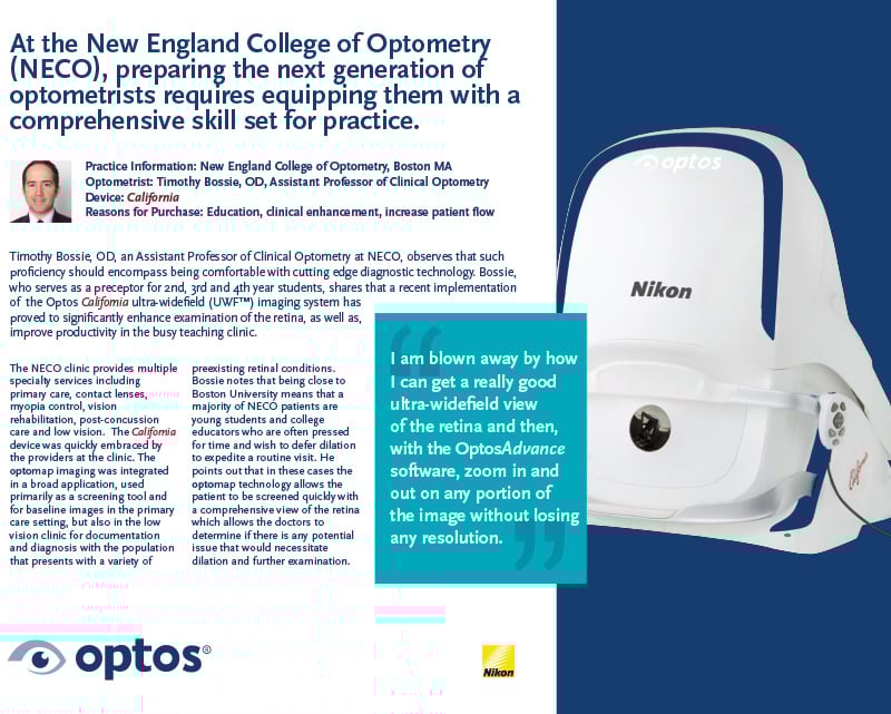
“Our providers and students are amazed out how far out in the periphery the optomap images can capture. I am blown away by how I can get a really good ultra-wide field view of the retina and then, with the OptosAdvance software, zoom in and out on any portion of the image without losing any resolution.” Quotes Bossie on the Optos California.
Over the years, optomap has proven to be irreplaceable in the NECO clinic and has aided both in efficient patient flow as well as a much-needed diagnostic and teaching tool. UWF imaging technology gives eye care providers imagery and diagnostic information about the retinal periphery that can’t be provided by small-field imaging methods. As an eyecare professional, you may increase your practice efficiency and enhance patient care with optomap, just like Dr. Bossie, and so many others. Contact us today to find out more.