According to the Centers for Disease Control and Prevention (CDC), thousands of people in the U.S. suffer eye injuries while on the job which will require medical attention. Each day, over 2,000 Americans suffer an eye injury. This means that almost one million Americans have experienced some vision loss due to eye injury. While most of these injuries are attributed to small particles like dust or wood chips hitting the eye, other injuries can result from a sharp object penetrating the eye or blunt force trauma, which can cause permanent vision loss or even the loss of an eye. Workers in other industries, such as health care, face the risk of coming in contact with an infectious disease if proper precautions aren’t taken.
March has been deemed Workplace Eye Wellness Month in order to shed light on preventable eye injuries and share some pointers for workers to keep their eyes safe while at work. Its often assumed that work-related eye injuries are isolated to outdoor jobs and those relating to physical labor but ironically, the most common eye problem in the workplace is computer vision syndrome.
Making routine eye exams a part of a yearly, preventative routine aid in the strides to prevent illness rather than treat it as it appears, but for many it is still not the norm. Many adults with 20/20 vision and no eye-related symptoms will often forgo an annual eye exam, while many ocular diseases are asymptomatic in early stages. Early detection of these diseases can have a significant impact on courses of treatment and the probability of positive outcomes.
Eyecare professionals can greatly enhance their comprehensive exams with the use of ultra-widefield (UWF™) optomap technology. optomap is specifically designed to provide an UWF image of the retina, and it is the only technology that captures 200-degrees of the retina a single capture and in less than ½ second. One illustration of how UWF imaging can improve the early detection of eye disease are the results of what amounts to an inadvertent experiment in the screening of healthy individuals. Training for Optos users and new Optos employees requires hands-on training on UWF imaging systems. Part of that involves trainees taking optomap images of themselves. These training exercises have revealed some surprising results:
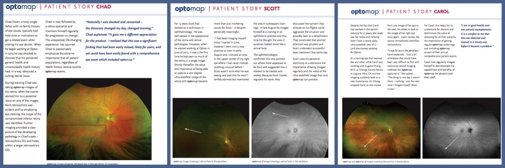
— Chad, who had no history of eye problems. Prior to coming to Optos his full schedule and absence of symptoms had made getting an optometric exam a low priority. During training Chad’s instructor observed faint retinoschisis on one of Chad’s optomap images. Further examination using eye steering enabled Chad and his instructor to fully visualize the extent of Chad’s retinoschisis, which included holes in a larger area of retinoschisis in his right eye. Chad’s now receiving regular medical care and follow-up.
— Scott’s initial instruction included routine color optomap images of his own eyes. Then, in early August, he imaged himself during a training session and noticed an area in his upper right eye that he’d not seen on earlier images. Unsure of what this was, he kept track of it during other training sessions. In late August Scott imaged himself again during a training session at an ophthalmic practice and this time the suspect area had resolved itself into something identifiable – a retinal hole. The ophthalmologist participating in the training confirmed this and also observed related leakage. He conferred with Scott and discussed the possible risks associated with his frequent business travel. Scott received laser treatment later that same day.
— Carol, an optical industry professional with 27 years of experience, had never viewed her retina nor had an ultra-widefield examination until she started working for Optos. Then, during a training session with an MD, the doctor stopped Carol as they were viewing color optomap images of her eyes. Asking to look once more at the image of her right eye, the doctor identified an area of retinoschisis visible on the far periphery of the retina. Carol has received follow-up treatment and makes a point during training sessions to image herself and to share her personal experience with the doctors and medical professionals with whom she works.
These stories are by no means unusual, and while not part of a statistically significant study, they make a simple, important statement about ocular healthcare – the wider use of ultra-widefield retinal imaging for routine screening has the potential to significantly improve personal health outcomes. Find an eye care professional who uses optomap, today!
Sources:
https://www.preventblindness.org/protect-your-vision-job
https://yoursightmatters.com/march-is-workplace-eye-wellness-month