As a private practice optometric physician and professor of optometry near Portland, Oregon, Lorne Yudcovitch, OD had the opportunity to experience optomap technology on a frequent basis for nearly a decade, both in the clinical setting and as a tool for instructing his students. While he valued the ultra-widefield view and the innovative capabilities, he did not purchase an Optos device until 2012.
“I felt that the optomap technology had improved not only in resolution, but it was more user-friendly, making it much easier to position the patient – and we loved the autofluorescence modality. That has become really invaluable.” Yudcovitch explained how optomap Daytona can quickly and easily reveal issues that might otherwise go unnoticed.
He describes a recent scenario when Lila, a 54-year-old Caucasian female, came to the clinic with night vision complaints. Yudcovitch shares that she was correctable to 20/20 vision in each eye and that he found no retinal abnormalities with conventional imaging and ophthalmoscopy. However, the optomap af image revealed significant hyper and hypo fluorescence mottling in the posterior pole in both eyes. Yudcovitch immediately referred her for further retinal examination and genetic testing which revealed a hereditary PRPH2 gene mutation variant.

optomap image of Lila’s right eye
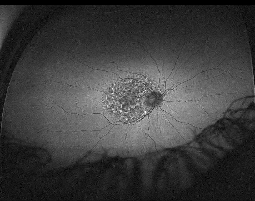
optomap af image of Lila’s right eye showing her genetic mutation
The gene product of PRPH2 is important to the integrity and stability of the structures that contain light-sensitive pigments. Slowly progressive loss of vision is typically seen in middle-aged individuals and the changes may be imperceptible initially, however, Autoflouresence can reveal abnormalities before patients become symptomatic.1 Yudcovitch acknowledges that optomap af was instrumental in the detection of the disease. Further genetic testing of the patient’s blood relatives was also recommended.
Prior to investing in optomap technology, Yudcovitch had relied on a retinal camera which was not utilized nearly as much because of the time and effort involved. The split-second, ease-of-use and dynamic modalities of optomap now enable them to image every patient in pre-test for documentation, as well as for detection of issues, like Lila’s, that could be otherwise missed.
Yudcovitch notes that patient response to the optomap experience has been favorable. “They find it extremely interesting and hi-tech. optomap has become very important in terms of patient education. It provides a good chronological assessment so that I can quickly and easily show my patients a side-by-side comparison and they can better understand what is occurring in their eyes. In fact, optomap has proven to be an excellent internal marketing tool because patients will often go and tell their family and friends about their experience and then these individuals start coming to see us.”
Ultimately, however, Yudcovitch places the greatest value on the enhanced quality of care for his patients. He emphasizes that optomap has helped the doctors at the practice to catch numerous sight and life-threatening pathologies from subtle hemorrhages and peripheral retinal tears to serious vascular issues and melanomas.
“Our Daytona is amazing. It does not replace what I do, but it enhances my abilities during the exam and that is invaluable. It’s definitely worth the cost and would move any practice to a higher level of care.”
As an eyecare professional, you may increase your practice efficiency and enhance patient care with optomap, just like Dr. Yudcovitch, and so many others. Contact us today to find out more.
1 https://disorders.eyes.arizona.edu/category/genes/prph2