November is Diabetes Awareness Month. In 2015, 30.3 million Americans, or 9.4% of the population, had diabetes and approximately 1.25 million American children and adults had type 1 diabetes. These numbers are on the rise and the disease manifests with deleterious and deadly impact throughout the body – including the eye. An understanding of the disease, early detection and treatment are more imperative than ever.
A recent study cooperatively funded by the National Eye Institute, the National Institute of Diabetes and Digestive and Kidney diseases and the US Department of Health and Human Services concluded that optomap ultra-widefield (UWF™) retinal imaging is a useful diagnostic tool for detection and assessment of severity of diabetic retinopathy (DR). The study published recently in JAMA Ophthalmology demonstrates that optomap UWF imaging can be used reliably in place of Early Treatment Diabetic Retinopathy Study (ETDRS) 7-Field imaging in clinical use and future clinical trials. The paper, which builds off recent single site studies that found moderate to perfect agreement between the modalities, supports these findings through data acquired over a two-year period from multiple sites.
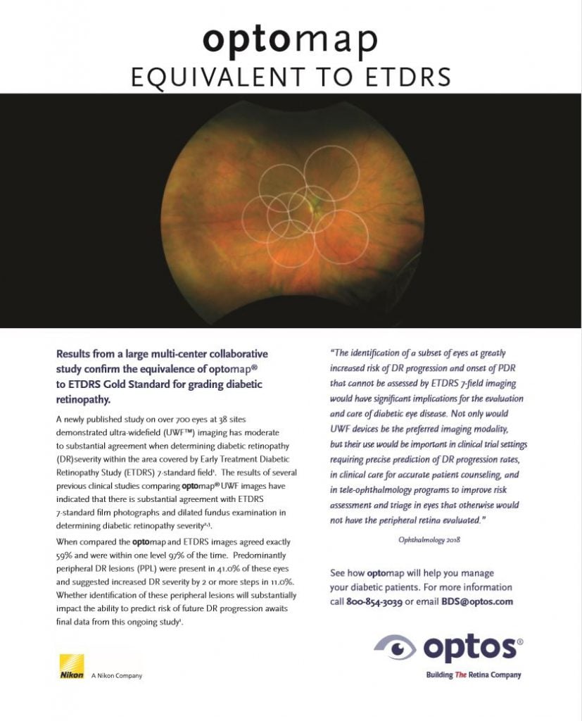
The gold standard assessment of DR severity has been based on grading of lesions within the ETDRS 7 standard fields. These 7 fields are time consuming in their acquisition, require dilation and once compiled represent only 34% of the retinal surface. Advances in retinal imaging technology now allow UWF imaging to capture 82% of the retina in a single image and in less than ½ second without the need for dilation. This collaborative study offers that given the technological advances now enabling UWF imaging, and the potential benefits of this approach, there may be substantial impetus for moving to UWF imaging if it is compatible in determining DR severity, and if pathology in the retinal periphery provides additional clinically useful information on prospective worsening of retinopathy.
In this current multi-site study, there were 737 gradable eyes on both ETDRS 7-field images and UWF images masked to contain the same 7 fields after adjudication; 59% had exact agreement, and 96% were within 1 step of agreement. The conclusion of the study clearly supported moderate to perfect agreement between modalities within the limited masked scope of the current gold standard. As seen was seen in previous studies, when the area outside of ETDRS was assessed predominantly peripheral DR lesions (PPL) were present in 41.0% of these eyes and suggested increased DR severity by 2 or more steps in 11.0%.
Cursorily referenced in this preliminary paper was a consideration of efficiency between modalities. This initial study release notes that the use of UWF imaging in clinical settings not only increases the frequency of DR identification nearly 2-fold but also reduces acquisition time by more than half, ungradable image rate by 71% and image evaluation by 28% compared with non-mydriatic fundus photography.
The study suggests the possibility of UWF imaging becoming a preferred method of assessment of DR severity, not only because of moderate to perfect agreement between modalities within the ETDRS scope, but also because of the information found in UWF outside the ETDRS mask. This raises the question regarding the potential for detecting DR change and severity earlier. Data collected from a previous study, suggests that lesions observed outside of the area captured within ETDRS may identify a possible subset of patients with the disease that may be more aggressive. These lesions were found to suggest a 4.7 times greater risk of worsening to treatable DR over a period of four years. That study also concludes that the identification of a subset of patients at greatly increased risk of experiencing DR progression and onset of proliferative DR that cannot be assessed by ETDRS 7-field imaging, would have important implications for the evaluation and care of diabetic eye disease.
The significance of the additional peripheral information gleaned through UWF imaging in assessing the risk of future DR progression will develop with the data collected from this ongoing study. The complete study and summary document, as well as our entire clinical library is available on our website. We encourage you to learn more about the clinical benefits of utilizing ultra-widefield optomap in your practice or clinic.
Sources:
Comparison of Early Treatment Diabetic Retinopathy Study Standard 7-Field Imaging with Ultra Widefield Imaging for Determining Severity of Diabetic Retinopathy. Journal of American Medicine, 2018
Peripheral Lesions Identified on Ultra Widefield Imaging Predict Increased Risk of Diabetic Retinopathy Progression Over Four Years. Ophthalmology 2015