Those beautiful lashes and bright eyes may have aesthetic appeal, but lashes and eyelids may obscure areas of interest within an optomap® image. Fortunately, patient lid and lash taping techniques can help eyecare professionals obtain unobstructed images for more effective analysis of their ultra-widefield (UWF™) retinal images.
Eyecare professionals use retinal imaging to identify and diagnose ocular and systemic diseases. Indicators of systemic disease and precursors to various eye conditions often develop first on the periphery of the retina. Current retinal examination methods provide a small field of view, thereby missing potentially important indicators. As the field of view becomes greater with optomap, artifacts such as lid and lash are captured in the image.
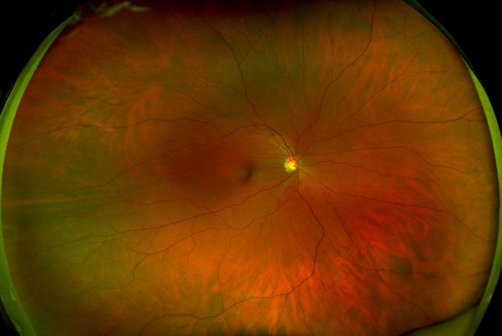
UWF color optomap image of a healthy retina with imperceptible lid and lash.
Lid and lash taping reduces artifacts to provide an unobstructed view of the entire retina, from the pole to the periphery. Lid and lash taping is not painful, but patients sometimes balk at the suggestion. Patient education about lid and lash taping helps alleviate concerns. Implementing a precise procedure for lid and lash taping, and performing retinal imaging immediately after taping, improves patient comfort.
Reducing Lashes in Images
An efficient taping procedure is essential in reducing lashes in retinal images. Taping should be fast, efficient, and performed with patient comfort in mind. Choice of tape is also important. A quality hyperallerginic latex-free micropore surgical tape that is very gentle on the skin and leaves minimal adhesive residue upon removal has been found to be successful.
Steps for reducing lashes in images when using a Daytona device, include:
- Set the tablet to “auto-capture,” which allows for quick image capture.
- Cut 1.5 inches of a piece of tape.
- Instruct the patient to look down, which lowers the upper eyelid.
- Holding the sticky side of the tape up, place the sticky side of the tape under the patient’s upper eyelashes.
- While the patient continues to look down, gently move the tape and lashes toward the eyebrow.
- Secure the tape to the skin of the patient’s forehead, as close to the brow as possible.
- Guide the patient to the aperture and begin imaging immediately.
- Remove the tape as quickly as possible after imaging to avoid patient discomfort.
- Gently remove the tape, using a nasal to temporal movement.
Reducing Lid in Images
Eyelids can also interfere with retinal imaging, and eyecare professionals can take steps to reduce the appearance of lids in retinal images.
Steps for reducing eyelid interference in images when using a Daytona device, include:
- Set the Capture tablet to “auto-capture”.
- Cut a piece tape, measuring about 2-inches; more or less may be necessary according to the patient’s anatomy.
- Instruct the patient to look down.
- Holding the sticky side of the tape toward the patient, place one end of the tape at the eyelash margin, otherwise known as the tarsal plate.
- Gently move the tape and lid toward the brow as the patient continues to look down.
- Secure the tape to the skin of the brow so that it holds the lid up.
- Guide the patient to the aperture and begin imaging immediately.
- Wipe the patient’s brow with an alcohol wipe to help the tape stick to the skin if the patient is perspiring or if the examination room is warm.
- Gently remove the tape with a nasal to temporal movement after imaging.
The procedures for eyelash taping and eyelid taping are quite similar but have one main difference: the tape goes over the lashes to attach the lashes to the brow in lash taping, while the tape goes behind the lashes to attach the lid to the brow in lid taping.
Patient lid and lash taping can greatly improve the results of optomap UWF retinal imaging, particularly in patients with droopy eyelids. By implementing lid and lash taping techniques, eyecare professionals can greatly improve retinal analysis and patient experience.
Do you have any suggestions for improving optomap images that you can share? Please email us or leave a comment!