As the number of patients with diabetes worldwide continues to skyrocket, Australia’s current population of 24.6 million people is not immune. In fact, 1.1 million individuals have diabetes. As diabetes increases so does the prevalence of Diabetic Retinopathy (DR) making it the leading cause of visual impairment and preventable blindness in working age people in Australia1.
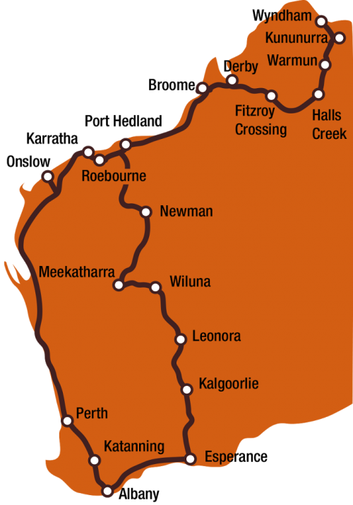
Lions Outback Vision in Western Australia Route Map
In general, most individuals are not aware how risky DR is to sight loss, therefore it is critical for them to be educated on the risks. Even if a patient is asymptomatic, they may have early non-proliferative stages of DR which typically shows progressive vascular changes within the retina which usually occurs before any change to vision. Once disease reaches the proliferative stage, vision loss can occur rapidly and can be permanent. Key symptoms of proliferative DR include new abnormal blood vessels on the retinal surface, these new blood vessels are weak and may bleed causing retinal damage or lead to vitreous hemorrhage; this may also be associated with the formation of fibrous scar tissue and can cause retinal detachments.
optomap® ultra-widefield (UWF™) retinal imaging assists eyecare professionals detect, manage, and treat the diseases associated with diabetes. In Australia, optomap UWF imaging provides a cornerstone for an evolving standard of care for the diagnosis, management and treatment of diabetic retinal eye disease. Dr. Dianne Pyliotis of Sydney comments that, “the peripheral retina is most likely where you are going to see early signs of non- proliferative DR, which is why the optomap technology is a ‘no-brainer’”.
As an eyecare professional, providing your patients with the best care practices is vital in monitoring the progression of DR. Patient education is very important in this stage – optomap technology allows eyecare professionals to view and discuss changes in the retina with their patients. Dr Pyliotis believes when a patient can see the changes (or lack thereof) within their retina it can give them the motivation they need to begin to make changes to their lifestyle.
UWF Imaging and Clinical Validation
UWF optomap retinal imaging is performed by a specially designed scanning laser ophthalmoscope (SLO) that generates a high-resolution digital image of 200° (or about 82 percent) of the retina, in a single capture. By comparison, conventional 7 standard field (7SF) ETDRS photographs produce a smaller view (approximately 100°or 30%) of the retina. The SLO simultaneously scans the retina using two low-power lasers (red and green) that enable high-resolution, colour imaging of retinal substructures. Additional UWF imaging modalities which are supported with the technology are; fluorescein angiography (FA), fundus autofluorescence (FAF), and indocyanine green chorioangiography (ICG).
Studies have compared optomap colour images of diabetic retinopathy with the clinical examination, seven-standard field photography2,3. The comparison of UWF with ETDRS has determined the sensitivity and specificity to be in perfect agreement additionally 50% of eyes were found to have predominantly peripheral lesions only visualized by UWF imaging. These lesions have been associated with a nearly 5 fold risk in the progression of DR to proliferative disease.3 optomap colour imaging has been validated in other studies finding sensitivity and specificity to be similar to traditional retinal photographs as well as providing additional information about the health of the peripheral retina. These benefits have been evaluated for DR, AMD, retinal breaks and tears, paediatric retinal disease, myopia, ocular oncology, inflammatory disease and a variety of vascular and inherited retinal disorders4. Validation for use of UWF imaging in telemedicine programs has found nearly double the rate of diabetes and a significant reduction in ungradeables5.
Using UWF Imaging for Detection and Treatment of Diabetic Eye Disease in Indigenous Australians
Indigenous Australians are nearly seven times more likely to develop diabetes than their non-indigenous counterparts6. As with other remote and/or underserved populations, education and availability continue to be common barriers to receiving quality care. Professor Hugh R. Taylor AC, of the University of Melbourne has created the Roadmap6 to close the gap for vision by 2020. He has highlighted 42 key competencies to close the gap for vision between indigenous and non-indigenous Australians, he says;
“One of the key objectives met was getting the Medicare item number for the MBS 715 health check and retinal examination for people with diabetes in primary care… it is a real game changer.”
In partnership with Lions Outback Vision in Western Australia, Optos is determined to combat these numbers, increase patient education, and assist in this endeavour to save the sight of these peoples. The Lions Vision Van consists of three consulting rooms, filled with specialist equipment including optomap devices and can provide comprehensive eye care for cataracts, trachoma, glaucoma and DR. Each year it completes two circuits of rural Western Australia helping save sight for thousands of Indigenous Australians living in rural and remote communities.
A study on DR in the Kimberley Region, Western Australia found that one in five indigenous people who were tested at the rural sight had DR7. Without the deployment of eyecare health coordinator these cases would have gone undetected and patients could have faced loss of sight.
To find out more about incorporating optomap into your practice or clinic please contact us.
Resources:
- Macular Disease Foundation, Australia
- Nonmydriatic Ultrawide Field Retinal Imaging Compared with Dilated Standard 7-Field 35-mm Photography and Retinal Specialist Examination for Evaluation of Diabetic Retinopathy.
- Peripheral Lesions Identified on Ultra-wideField Imaging Predict Increased Risk of Diabetic Retinopathy Progression over 4 Years. Ophthalmology 2015.
- Nagiel et al. Ultra Widefield Fundus Imaging, A Review of Clinical Applications and Future Trends, Retina 2016.
- Silva. Identification of Diabetic retinopathy and Ungradable Image rate with Ultra-widefield Imaging in a national Teleophthalmology program. Ophthalmology. 2016.
- Vision 2020, Close the gap for vision. University of Melbourne April 2013.