Optometric practices face growing and more complex patient loads as they support the special needs of an aging population, an increase in diabetic patients and the complications associated with their condition, and manage patient flow so to strike a balance between patient experience and staff productivity. Many optometrists are satisfying all of these demands by incorporating ultra-widefield (UWF™) retinal imaging into their practices.
Make optomap part of your comprehensive eye exam.
About Ultra-Widefield Retinal Imaging
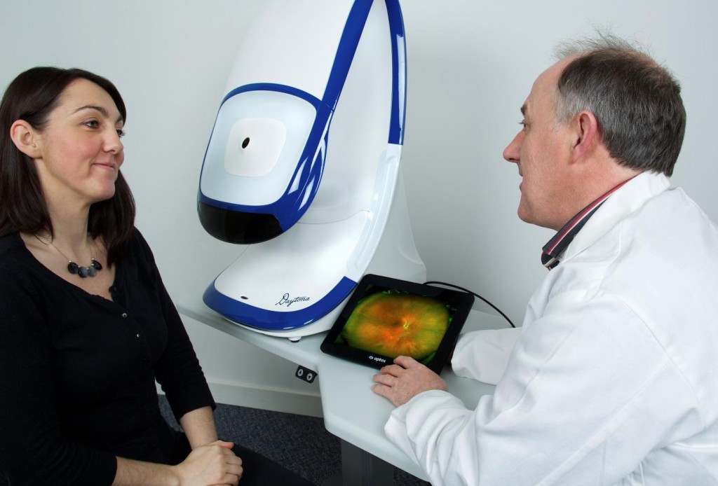
UWF retinal imaging is performed by a specially designed, table-top scanning laser ophthalmoscope (SLO) that generates a high-resolution digital image covering 200° (or about 82%) of the retina. (this compares to other imaging techniques with fields of view typically under 45°).
The SLO simultaneously scans the retina using two low-power lasers (red and green) that enable high-resolution, color imaging of retinal substructures. The resulting UWF high resolution digital image – the optomap – is produced in a single capture without pupil dilation. Tabletop systems designed for optometric practice (Daytona) provide both UWF color imaging and UWF autofluorescence modalities in a single scan.
Unlike routine slit lamp examinations, optomap can be performed by a trained technician. The image is captured in less than a second using automatic prompts that position the patient for an accurately. The system can image through pupils as small as 2 mm and through many cataracts.
Improved Patient Experience
UWF retinal imaging impacts the patient experience in ways that help redefine what it is to visit the optometrist:
— Pupil dilation is usually not needed, significantly shortening the visit and eliminating a sometimes uncomfortable process. This can also encourage patients to make and keep follow-up visits.
— For patients that are difficult to image – like children – UWF imaging can transform what is to them an extended, difficult and upsetting procedure into something more akin to a game.
— Practitioners using optomap during patient discussions are able to illustrate their findings with credible, visually compelling images. optomap images are ideal for showing the presence or progression of pathologies, creating added impetus for patients to follow through on recommended referrals.
It’s for these reasons many optometric practices are now starting to publicize their use of UWF imaging on their web sites and in other patient communications. Optometrists using UWF imaging report that many patients are happy to personally cover the cost of including optomap in their comprehensive exam in order to shorten their exam, avoid dilation and create a permanent record.
A Better Diagnostic Process
UWF imaging improves the diagnostic process by separating examination and diagnostic activity. Where slit lamp examination requires the practitioner to multi-task – perform the exam, evaluate what appears in the field of view, possibly take photographs, and stay engaged with their patient – optomap allows the practitioner to perform an offline and uninterrupted evaluation of the patient’s examination results. Evaluative support software can also be used to support the diagnostic evaluation.
Productivity and Workflow Gains
One only has to look at an optometrist’s waiting room and the patients waiting for their pupils to fully dilate to appreciate the positive effect UWF imaging can have on patient flow. The process of guiding patients in and out of examining rooms during a dilated eye examination consumes space and staff resources better applied to high value tasks.
Personal productivity is also improved when UWF imaging is utilized in optometric practice. UWF images can be performed by a trained technician or nurse, obviating the need for a routine slit lamp examination and freeing up optometrist time. Further, the optometrist who evaluates a patient’s optomap (current and prior) before examining the patient, can use this to help plan their examination and discussion – improving their productivity while enabling more meaningful patient engagement.
Office workflow is streamlined with UWF imaging. optomap images are easily appended to patient EHRs (electronic health records), and workflow software can be used to make referrals or even request second opinions. The inclusion of optomap images in referrals is another benefit. They can simplify the referral process while improving the quality and precision of the diagnostic information being passed on to the referring physician.
Adding UWF retinal imaging capability to an optometric practice is an important decision, but there may be no other single investment practitioners can make that can simultaneously improve the patient experience, enhance the diagnostic process, and increase office and staff productivity. For examples of how your peers are benefiting by incorporating optomap into their practices, please contact us.