optomap for telemedicine programs
More Patients, Not Enough Vision Care Providers
There are an estimated 415 million people in the world with diabetes. That population will climb to 642 million by 2040. The 104 million diabetes patients now in North America and Europe will grow at a slower rate, but by 2040 will still total over 132 million people.
The public health issues presented by diabetes are numerous, but vision care is of particular concern. Some level of diabetic macular edema (DME). Complicating the picture is the high rate of undiagnosed diabetes – it’s estimated at over 27% of cases in the US. Finally, early stage DR is often asymptomatic, giving patients no reason to seek vision care.
Access is another part of the problem. It’s no surprise that it can be difficult to receive proper vision care in the developing world, but there are similar issues in developed nations. A 2011 study found that 24% of the counties in the United States had no optometrists of ophthalmologists. More than 60% of counties were ranked in the lower two quartiles for vision care provider availability1.
Expanding Diabetic Retinopathy Screening
A strategy to make DR screening more widely available is the subject of a recent paper2. Rather than advocating the use of existing methods, the paper shows how new technology combined with telemedicine can improve health outcomes.
Cost and access drive the need for better screening technology. The current standard for managing diagnosed DR is ETDRS seven-field color fundus photography (7SF). Processing 7SF imagery normally requires an ophthalmic photographer, a grader to evaluate the images, and a medical professional to assess the results. The procedure, performed in a doctor’s office, is costly and not very portable.
The authors suggest that effective DR screening technologies should have the ability to be easily deployed to remote locations; do no more than accurately identify patients who need to be referred to a specialist, and; reduce the overall costs of testing and treatment. Ideally, screening should be conducted without dilation to cut examination time and reduce the risk of angle-closure glaucoma.
The Ultra-widefield Imaging Solution
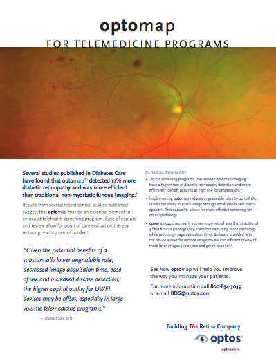
A growing body of clinical evidence is validating ultra-widefield (UWF™) imaging as an effective DR screening technology. UWF optomap® color imaging is typically performed without dilation and can be performed by a clinical nurse or technician.
— One study covering a large telemedicine program compared the gradability of UWF images compared to multi-field fundus images taken without dilation. UWF imagery was ungradable in only three to four percent of exams versus multi-field fundus images that were ungradable in twenty-seven percent of exams3.
— optomap retinal imaging delivers diagnostic results that are equivalent to the ETDRS gold standard.
“Nonmydriatic ultrawidefield images compare favorably with dilated ETDRS photography and dilated fundus examination (by a retina specialist) in determining DR and DME severity4 . . .”
UWF optomap retinal imaging can fulfill diagnostic and operating requirements for a robust DR screening solution.
The Public Health Impact
Numerous studies have quantified how telemedicine programs can improve DR screening rates and ocular health outcomes. One telemedicine program of the US Indian Health Service reported a 50% increase in annual retinal exams5. Another study found that participants in a telemedicine program were up to two times more likely to get a comprehensive DR exam than a group being monitored using small field fundus photography methods6.
Proven screening technologies like UWF imaging are building confidence that telemedicine can have a major, positive impact on DR and DME treatment. The task ahead? Public health authorities, private insurers and practitioners need to widen access to telemedicine and establish delivery methods that optimize cost-effectiveness. The result will be important public health gains that will benefit tens of millions of people around the globe.
About optomap and UWF Imaging
UWF imaging technology gives ocular health practitioners imagery and diagnostic information about the retinal periphery that can’t be provided by conventional imaging methods. Starting with color (red and green) optomap imaging, Optos has systematically extended its UWF-based technology into a multi-modal platform that supports fundus autofluorescence (optomap af), fluorescein angiography (optomap fa) and indocyanine green angiography (optomap icg).

Sources:
- Gibson DM. The geographic distribution of eye care providers in the United States: Implications for a national strategy to improve vision health. Prev Med. 2015;73:30–36.
- Gupta A, Cavallerano J, Sun JK, Silva PS, Evidence for Telemedicine for Diabetic Retinal Disease, Seminars in Ophthalmology. 2016 Oct 17:1-7.
- Silva PS, Horton MB, Clary D, et al. Identification of diabetic retinopathy and ungradable image rate with ultrawide field imaging in a national teleophthalmology program. Ophthalmology 2016;123(6):1360–1367
- Silva PS, Cavallerano JD, Sun JK, Noble J, Aiello LM, Aiello LP, Nonmydriatic Ultrawide Field Retinal Imaging Compared with Dilated Standard 7-Field 35-mm Photography and Retinal Specialist Examination for Evaluation of Diabetic Retinopathy, American Journal of Ophthalmology, May 2012
- Wilson C, Horton M, Cavallerano J, Aiello LM. Addition of primary care-based retinal imaging technology to an existing eye care professional referral program increased the rate of surveillance and treatment of diabetic retinopathy. Diabetes Care 2005;28(2):318–322.
- Mansberger SL, Sheppler C, Barker G, et al. Long-term comparative effectiveness of telemedicine in providing diabetic retinopathy screening examinations: A randomized clinical trial. JAMA Ophthalmology 2015;133(5):518–525.