Blurred vision in one or both eyes can be a sign of a simple change in vision or something much more serious, especially when it occurs suddenly and persists for a few days. A 36-year-old new patient of Dr. Paula Koch, OD, noticed a “blob-like, blurry-ness” in his left eye and knew a visit to the eye doctor was necessary to get to the bottom of the issue.
While the patient’s medical history was unremarkable, his ocular history included a high keratoconic prescription, partial albinism, and amblyopia. Based on his ocular history and current symptoms, Dr. Koch recommended an optomap exam. The patient agreed to the exam, which revealed a rhegmatogenous retinal detachment spanning the 11-to 7-o’clock region of the nasal retina OS, as well as a small tear at 9 o’clock and some chorioretinal scarring in the far periphery and Weiss’ ring inferior to the disc. Because of the patient’s partial albinism and other unusual aspects of the optomap’s finding, including the blister-like appearance of the detachment, Dr. Koch referred the patient to a retinal specialist within 20 minutes of the optomap exam.
The retinal specialist performed a pneumatic retinopexy, which was unsuccessful and lead to a sclera buckle procedure. The procedure was a success and the patient returned to Dr. Koch for follow-up care. New optomap images were taken and Dr. Koch was able to compare them with the previous images and explain what had happened with the patient’s eyes and verify how efficient the specialist’s treatment was. The patient was fitted with new gas permeable lenses and his visual acuity is now 20/60.
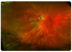
optomap images revealed rhegmatogenous retinal detachment.
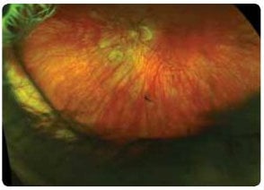
The patient’s eye in an optomap image taken after the sclera buckle procedure.
Dr. Koch credits the optomap with revealing a condition that could have affected the patient’s vision much more severely if it had not been diagnosed until later. Because of the optomap ultra-widefield image, she was able to give her patient a quicker, more accurate diagnosis, which helped save the vision of his left eye. Dr. Koch also shared that her patient trusts her with his vision and believes in the value of an optomap as a part of his annual eye exam.
This is just one of many instances in which an optomap exam helped save a patient’s vision. Please visit our website to read of other case studies.
As March is Save Your Vision Month, use this time to encourage patients to include an optomap as a part of their annual eye exam. With optomap’s ultra-widefield technology, conditions that could otherwise go unnoticed may be revealed, saving your patient’s vision and preserving it for years to come.